
Tympanoplasty and Middle Ear Surgery
The ear consists of three parts; external, middle and inner. Middle ear includes , ear drum and ossicles. gristle and ear ossicles. Any disease that affects the eardrum or ossicles can lead to conduction hearing loss by preventing sound from being transmitted to the inner ear from the external ear. Such a disease can range from a hole in the eardrum, to the destruction of one or more of the ear ossicles, to the disruption of the ossicular chain. When an inflammation develops in the middle ear, the ear can be perforated and the inflammation can flow out. This hole often heals itself and closes. If it does not improve, hearing loss usually occurs with intermittent or continuous ear flow and tinnitus.
Ear Care
You should not get water into your external ear channel. When bathing or washing your head, you should put a piece of cotton coated with vaselin jelly. When swimming, there is a benefit of tightly swimming over the petals with vaselin jelly. In addition, earplugs are sold in various sizes in the market and pharmacies.
You should avoid strong burning your nose. This event causes the flora of nose to reach the middle ear by means of the “Eustachian tube” between the nose and the middle ear. The nasal discharge should be drawn in and spit out. If you need to blow your nose so much, it is advisable to do this without closing your other nostril. As long as there is an ear flow, the ear should be apirated as much as possible without getting stuck inside. Drugs should be used if ear infections are present or have started. In the ear path, cotton can be put in order to determine the presence of the current, but this should not always block the front of the flow. Medical TreatmentDrug therapy will often stop the ear flow. Treatment requires intermittent cleaning of the ear, use of drops or dust. Certain people need oral antibiotics.
Surgical Treatment
The main purpose of the chronic middle ear surgery was to prevent harmful effects and inflammation for many years. Recently, methods have been developed to regain hearing.Many tissues can be used to patch or reconstruct the eardrum. These tissues are like “the skin of the ear canal, the membrane covering the ear, cartilage”. Damaged middle ear ossicles can be used for restorating ossicular chain to regain hearing. Sometimes, instead of a ossicle, cartilage or prosthesis can be used. The middle ear is a hollow chamber in the bone of the skull. It is separated from the outside world by a thin membrane about half-an-inch in diameter, the eardrum. The middle ear area is lined by the same kind of mucous membrane that lines nose and mouth. It is connected to the back of the nose, just above the soft upper portion of the mouth, by a narrow passage called the eustachian tube.
The eustachian tube lies closed until the swallowing movement pulls it open and allows fresh air to enter the middle ear. The fresh air is needed to replace oxygen that has been absorbed by the middle ear lining. The fresh air equalizes the middle ear pressure with the air pressure outside the head. Some people hear this burst of fresh air as a pop or click.
Suspended within the middle ear is a chain of three small bones, the ossicles, which conduct sound vibrations from the eardrum across the middle ear into the fluid-filled inner ear. Inside the inner ear these vibrations are converted to nerve signals that are carried by the auditory nerve to the brain.
The mastoid bone is an extension of the air space of the middle ear. It is made up of small interconnected air spaces similar to a honeycomb. Its function is not clear, but it is often involved in chronic ear infections. Within it lie the structures of the inner ear responsible for balance and facial expression.
What is Chronic Otitis Media?
Chronic Otitis Media (COM) is the term used to describe a variety of signs, symptoms, and physical findings that result from the long-term damage to the middle ear by infection an inflammation. This includes the following:
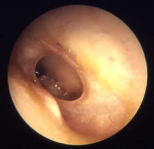
- Severe retraction or perforation of the eardrum (a hole in the eardrum)
- Scarring or erosion of the small, sound conducting bones of the middle ear
- Chronic or recurring drainage from the ear
- Inflammation causing erosion of the bony cover or the facial nerve, balance canals, or cochlea (hearing organ)
- Erosion of the bony borders of the middle ear or mastoid, resulting in infection spreading to the meninges (the coverings of the brain) or brain
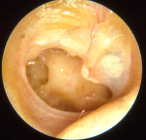
- Presence of cholesteatoma
- Persistence of fluid behind an intact eardrum
- How Does Chronic Otitis Media Occur
- The eustachian tube becomes blocked by swelling or congestion in the nose and throat, by swelling of the mucous membrane in the middle ear, or by swelling of the mucous membrane of the eustachian tube itself, the air pressure in the middle ear cannot equalize properly. A negative pressure develops, and if the obstruction is prolonged, fluid may be drawn into the air space of the middle ear from the mucosa. This may occur with a cold or flu virus and is a common cause of ear infections in children (serous otitis media). Serous otitis media usually resolves without treatment, but may require a course of antibiotics or steroids. It is a common reason for placement of tubes in children and adults.
- If the eustachian tube blockage persists, chronic changes in the tissue of the middle ear begin to occur. First, the mucous secretions become thicker, and therefore less likely to drain. Then the membranes themselves begin to thicken and become inflamed. The defense mechanisms of the eustachian tube and middle ear become compromised and bacteria normally present in the nose may enter the middle ear and cause a painful condition called acute otitis media. This responds to antibiotic treatment, but may require placement of tubes.
- The negative pressure in the middle ear or alternating periods of negative, normal and positive pressure may deform the eardrum. In the long term, the eardrum may become severely distorted, thinned, or even perforated. These changes may cause hearing loss and a sensation of pressure. When there is a hole in the eardrum, the natural protection of the middle ear from the environment is lost. Water and bacteria entering the middle ear from the ear canal can cause inflammation and infection. Drainage from the ear is a sign of a perforation.
- Inflammation and infection in time can cause erosion of the ossicles and the walls of the middle and inner ear. The patient may experience hearing loss, imbalance, or weakness of facial movement on the affected side. In rare instances, the infection may extend deeper into the head, causing meningitis or brain abscess.
- A cholesteatoma, or skin cyst, is essentially skin in the wrong place. Epidermal skin from the ear canal or outside surface of the eardrum, like that on the back of the hand, does not belong in the middle ear. If it is trapped by a deformed eardrum or migrates through a perforation, it tends to grow out of control and can cause significant damage to the structures of the middle ear and mastoid.
How Do I Know If I Have Chronic Otitis Media?
Warning signs of chronic otitis media include:
- Persistent blockage of fullness of the ear
- Hearing loss
- Chronic ear drainage
- Development of balance problems
- Facial weakness
- Persistent deep ear pain or headache
- Fever
- Confusion or sleepiness
- Drainage or swelling behind the ear
Chronic otitis media generally occurs gradually over many years in patients with longstanding or frequent ear trouble. However, it can occasionally develop over several months in a patient with no previous history of ear disease. Any of the above symptoms should prompt an evaluation by an ENT or otologist/neurotologist.
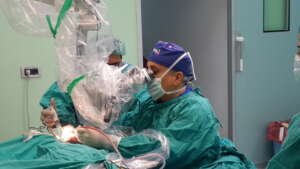
Tympanoplasty
Tympanoplasty is the repair of the eardrum and/or middle ear bones. Tympanoplasty is most commonly performed for repair of the eardrum. Eardrum perforations (holes) are usually caused by trauma or infection. Two methods are commonly done to repair the eardrum.
The first is a Myringoplasty which is used to repair a small hole in the eardrum. In this operation, the hole’s margin is rimmed, a process which removes skin and tissue, and a small piece of fat is placed into the hole. This operation does not take a long time and in adults can be performed in an office setting under local anesthesia.
The second operation is a formal Tympaoplasty. In this operation, the middle ear is entered through a canal skin flap and a piece of muscle tendon or fascia is placed beneath the perforation. This operation is usually performed in the operating room and takes much longer under a general anesthesia.
Mastoidectomy
Mastoidectomy is a surgical procedure to remove an infected portion of the bone behind the ear when medical treatment is not effective. This surgery is rarely needed today because of the widespread use of antibiotics.
Purpose
Mastoidectomy is performed to remove infected air cells within the mastoid bone caused by mastoiditis, ear infection, or an inflammatory disease of the middle ear (cholesteatoma). The cells are open spaces containing air that are located throughout the mastoid bone. They are connected to a cavity in the upper part of the bone, which is in turn connected to the middle ear. As a result, infections in the middle ear can sometimes spread through the mastoid bone. When antibiotics cannot clear this infection, it may be necessary to remove the infected air cells by surgery. Mastoidectomies are also performed sometimes to repair paralyzed facial nerves.
Description
Mastoidectomy is performed less often today because of the widespread use of antibiotics to treat ear infections.
There are several different types of mastoidectomy:
• Simple (or closed). The operation is performed through the ear or through a cut (incision) behind the ear. The surgeon opens the mastoid bone and removes the infected air cells. The eardrum is cut (incised) to drain the middle ear. Topical antibiotics are then placed in the ear.
• Radical mastoidectomy. The eardrum and most middle ear structures are removed, but the innermost small bone (the stapes) is left behind so that a hearing aid can be used later to offset the hearing loss.
• Modified radical mastoidectomy. The eardrum and the middle ear structures are saved, which allows for better hearing than is possible after a radical operation.
The wound is then stitched up around a drainage tube, which is removed a day or two later. The procedure usually takes between two and three hours.
Aftercare
Painkillers are usually needed for the first day or two after the operation. The patient should drink fluids freely. After the stitches are removed, the bulky mastoid dressing can be replaced with a smaller dressing if the ear is still draining. The patient is given antibiotics for several days.
The patient should tell the doctor if any of the following symptoms occur:
• Bright red blood on the dressing.
• Stiff neck or disorientation. These may be signs of meningitis.
• Facial paralysis, drooping mouth, or problems swallowing.
Risks
Complications do not often occur, but they may include:
• Persistent ear drainage.
• Infections, including meningitis or brain abscesses.
• Hearing loss.
• Facial nerve injury. This is a rare complication.
• Temporary dizziness.
• Temporary loss of taste on the side of the tongue.
Cholesteatoma
A rare but chronic inflammatory disease in which skin cells and debris collect in the middle ear, usually as a result of an ear infection.
Mastoiditis
An inflammation of the bone behind the ear (the mastoid bone) caused by an infection spreading from the middle ear to the cavity in the mastoid bone.
Orta Kulak ve Kulak Zarı Ameliyatları
Kulak, dış, orta ve iç olmak üzere üç bölümden oluşur. Orta kulak, kulak zarı ve kulak kemikçiklerini içerir. Kulak zarını veya kemikçikleri etkileyen herhangi bir hastalık, sesin dış kulaktan iç kulağa iletilmesine engel çıkartarak iletim tipi işitme kaybına yol açar. Böyle bir hastalık, kulak zarındaki bir delikten, kulak kemikçiklerinin bir veya daha fazlasının tahribinden, kemik zincirinin bozulmasına kadar değişik çerçevede olabilir.
Orta kulakta bir iltihap oluştuğu zaman, kulak zarı delinerek iltihap dışarı akabilir. Bu delik, sıklıkla kendi kendine iyileşip kapanır. Şayet iyileşemezse, çoğunlukla kulak çınlaması ve aralıklı veya sürekli akıntının görüldüğü işitme kaybı oluşur.
Kulak Bakımı
Kulak zarınız delikse kulağınızın içine su kaçırmamalısınız. Banyo yaparken veya başınızı yıkarken kulağınıza vazelinle sıvanmış bir pamuk parçası koymanız gerekir. Yüzerken, vazelinli pamuğun üzerine sıkı sıkı yüzme beresi geçirmenizde fayda vardır. Ayrıca market ve eczanelerde değişik boyutlarda kulak tıkaçları da satılmaktadır.
Burnunuzu kuvvetli sümkürmekten kaçınmalısınız. Bu olay, geniz ile orta kulak arasında bulunan “östaki borusu” vasıtası ile burun ve genizdeki mikropların, orta kulağa ulaşmalarına sebep olur. Burun akıntısı içe çekilmeli, tükürülmelidir. Burnunuzu sümkürmeniz çok gerekiyorsa, bunu diğer burun deliğinizi kapatmadan yapmanız tavsiye edilir.
Kulak akıntısı olduğu müddetçe, kulak, mümkün olduğunca içerisine fazla bir şey sokulmadan temizlenmelidir. Kulak akıntısı varsa veya başlamışsa ilaç kullanılmalıdır. Kulak yoluna, akıntının varlığını tespit etmek amacı ile pamuk konabilir fakat bu, akıntının önünü sürekli tıkamamalıdır.
İlaç Tedavisi
İlaç tedavisi çoğunlukla kulak akıntısını durduracaktır. Tedavi, kulağın aralıklı olarak temizlenmesini, damla veya toz kullanılmasını gerektirir. Belli kişilerde ağızdan antibiyotik kullanılması gerekir.
Cerrahi Tedavi
Yıllarca, kronik orta kulak cerrahisinin esas amacı, iltihabı kontrol altına almak ve geliştirebileceği zararlı etkilerini önlemek amacı taşımıştır. Son zamanlarda işitmenin de yeniden kazanılması için yöntemler geliştirilmiştir.
Kulak zarını yamamak veya yeniden yapmak için birçok doku kullanılabilmektedir. Bu dokular, “kulak kanalının derisi, kulak üzerindeki kası kaplayan zar, kıkırdak” gib
idir. Hasar görmüş bir kulak kemikçiği, yeniden yerleştirilebileceği gibi, yapma bir kemik de kullanılabilir. Bazen erimiş bir kulak kemikçiği yerine, kıkırdak da kullanılabilir.
Endoskopik Kulak Zarı Tamiri (Miringoplasti)
Çoğu orta kulak iltihabı kendi kendine iyileşir, bazıları da geride delik bir kulak zarı bırakır.
Kulak zarı tamiri orta kulağı korur ve bazen işitmenin de düzelmesini sağlar. Bu operasyon orta kulak kemikçiklerinde bir hasar olmadığı ve iltihabın kurumuş olduğu kişilerde yapılabilir. Operasyon, dış kulak yolundan veya kulak kepçesinin arkasından yapılır. Artık endoskopların yardımı ile, ciltte kesi yapmadan endoskopik olarak bu cerrahiler yapılmaktadır. Yukarıda belirtilen dokular, kulak zarı oluşturmak veya yama yapmak için kullanılır.
Kişi, bir gün içerisinde işine dönebilir. Tam iyileşme, yaklaşık altı hafta içerisinde olur ve ancak bu sırada operasyonun işitmeyi nasıl etkilediği anlaşılır.
Orta Kulak Tamiri (Timpanoplasti)
Orta kulak iltihabı, kulak zarıyla beraber orta kulak kemikçiklerinde de hasar meydana getirebilir. Timpanoplasti, hem kulak zarının hem de bu kemikçik zincirinin tamirine verilen isimdir. Bu operasyon, kulak zarının onarılmasını ve çoğu zaman işitmenin de daha iyi olmasını sağlar.
Operasyon, kulak kepçesi arkasından veya dış kulak yolundan yapılır. Orta kulak kemikçik zinciri, kemikçiklere yeniden şekil verilerek veya diğer dokularla onarılır, aynı zamanda zar tamiri de yapılır.
Bazı kişilerde, hem kemikçik zinciri hem de zarı aynı anda tamir etmek mümkün olmaz. Bu durumda önce kulak zarı onarılır, dört ay veya daha uzun bir süre sonra da kemikçik tamiri yapılır. Buna “aşamalı operasyon” adı verilir
Operasyon, genel anestezi altında gerçekleştirilir. Kişi, operasyondan sonra yedi ile on gün içerisinde işine dönebilir. İyileşme, yaklaşık altı haftada tam olur. İşitmedeki iyileşme birkaç ay hissedilmeyebilir.
Kulak Arkası Kemik (Mastoidektomi) ve Orta Kulak Tamiri (Timpanoplasti)
İltihap, bazı kişilerde, dış kulak yolundaki dokunun delik kulak zarından içeri girerek orta kulak ve kulak arkası kemiklerin içine yayılmasına sebep olur. Bu gerçekleşirse, deri ile kaplı, “kolesteatom” denilen bir kitle oluşur. Bu kitle, yıllar boyunca giderek genişler ve çevre kemik dokuları tahrip eder. “Kolesteatom” mevcutsa, kulak akıntısı daha sürekli ve sık, kötü kokulu bir hâl alır. Birçok hastada, sürekli olan akıntının sebebi, kulağı çevreleyen kemik dokunun kronik iltihabıdır.
Kolesteatom veya kemik dokuda bir iltihap oluşmuşsa, bu hastalığın artık ilaçla tedavisi şansı çok düşüktür. Damla veya ağızdan alınan anitibiyotikler, sadece geçici bir süre için iyileşme sağlarlar. Tedavi kesildikten sonra, tekrarlama sıktır.
Kolesteatom veya kronik kulak iltihabı, rahatsızlık veren akıntı ve işitme kaybı dışında bir şikayet yaratmadan, yıllarca öylece kalabilir. Fakat, lokal ilerleme ve basınç yaparak etrafındaki dokuya yayılması daha sıktır. Böyle olursa, hasta, sıklıkla kulakta dolgunluk ve künt bir ağrı hisseder. Şiddetli başdönmesi, yüz sinirinde zayıflık veya yüz felci gelişebilir. Bu şikayetlerden herhangi biri gelişirse, hasta, mutlaka cerrahi tedaviye alınmalıdır. Cerrahi, iltihabın ortadan kaldırılması ve gelişebilecek ciddi komplikasyonların önlenmesi için gerekli olur.
Kolesteatom, kulak kemiğinde önemli derecede hasar meydana getirmişse, cerrahi olarak bunu temizlemek zor olabilir. Operasyon, kulak arkasından bir kesi ile yapılır. Ana amaç, iltihabı ortadan kaldırmak ve kuru, emin bir kulak elde etmektir.
Kolesteatomlu hastaların yaklaşık üçte birinde, tek operasyonda hem iltihabı ortadan kaldırıp hem de işitmeyi onarmak mümkün değildir. İlk operasyonda iltihap ortadan kaldırılıp, kulak zarı tamiri gerçekleştirilebilir. Hasta, genellikle iki hafta sonra işine dönebilir.
İşitmeyi onarmak için ikinci operasyon altı ile yirmidört ay sonra yapılabilir.
Orta Kulak Tamiri (Timpanoplasti) ve Kulak Arkası Kemik (Mastoidektomi) Operasyonu Tekrarı
Bu operasyonun amacı, radikal kulak operasyonu sonucu oluşan boşluğun akıntısından kurtulmak, boşluğu doldurmak ve işitmeyi iyileştirmektir
Operasyon, kulak arkasından gerçekleştirilir. Kulak arkasındaki yağ veya kas, mastoid boşluğunu doldurmak için kullanılabilir. Mümkün olursa, kulak kemikçikleri, yapay kemikçikler veya kıkırdak, işitme mekanizmasını onarmak için kullanılabilir, fakat işitmede iyileşmeyi sağlamak için genellikle başka bir operasyon gerekir.
Operasyon, genel anestezi altında gerçekleştirilir. Hasta, operasyondan iki hafta sonra işine dönebilir. İşitmedeki iyileşme birkaç ay hissedilemeyebilir.
Radikal Mastoid Operasyonu
Bu operasyonun amacı, işitmeyi dikkate almadan, kulak iltihabının ortadan kaldırılmasıdır. Operasyon, çok inatçı kulak iltihabı olan hastalarda yapılır. Başlangıçta, orta kulak tamiri için uygun görülen hastalarda, nadiren de olsa radikal kulak operasyonu gerekli olabilir. Bu karar, operasyon esnasında verilmelidir. Kulak arkasından alınan bir kas nakli de, iyileşmenin daha düzgün olması için gerekli olabilir. Operasyon, genel anestezi altında gerçekleştirilir Hasta, genellikle iki hafta sonra işine dönebilir.
Mastoid Obliterasyon (Doldurma) Operasyonu
Bu operasyonun amacı, mastoid içerisindeki iltihabın kurutulması ve daha önce oluşturulmuş olan boşluğun doldurulmasıdır. İşitmedeki iyileşme dikkate alınmaz.
Operasyon, kulak arkasından bir kesi ile gerçekleştirilir. Mastoid boşluğu doldurmak için, kulak arkasından alınan kas veya yağ kullanılır. Operasyon, genel anestezi altında gerçekleştirilir. Hasta, işine iki hafta sonra dönebilir.
Sizin Kulağınızdaki Bulgular
Kulağınızdaki bulgular, muayene ve tetkiklerden sonra hekiminiz tarafından size açıklanacaktır.
Bunun sonucu nasıl bir operasyon gerektiğine birlikte karar verilecektir.
Genel Yorumlar
Şayet cerrahi başarısız olursa, işitme çoğunlukla operasyondan önceki seviyesinde kalır. Opere edilen hastaların yüzde üçünde, işitmenin daha da azalma ihtimali vardır. Operasyonu takiben, nadiren, bir süre, kulak akıntısı, başta uğultu, tat bozukluğu veya sersemlik hali olabilir. Hastaların yüzde birinden daha azında, yüzde zayıflık gelişebilir. Bu çoğunlukla geçici bir komplikasyondur.
Şayet şu an operasyon yapılmazsa, yıllık kontrollerinizi yaptırmanız, özellikle kulak akıntısında hemen muayeneye gelmeniz önerilir. Şayet kulak içerisinde veya etrafında künt bir ağrı, akıntıda artma, baş dönmesi, yüzde uyuşma-kayma hali gelişirse, hemen doktorunuza başvurmalısınız.